The Ligand Binding Pocket
The ligand binding pocket of k-ORs was seen to have a mixture of unique and more common structural properties. The k-OR ligand binding pocket was seen to have an Asp138 deep in the cavity, which formed an ionic interaction with the bound JDTic ligand. This residue is present in most aminergic GPCRs and opioid receptors and provides a contact for stabilizing interactions with a protonated amine group on the ligand. Along with the ECL2 β-hairpin structure covering the binding pocket, this Asp138 supposes the main shared feature (with chemokines and aminergic receptor families) in the binding pocket of the k-OR. The binding pocket was narrower and deeper (see Figures 5 and 6) than that of closely related proteins such as CXCR4 and was also seen to differ significantly in the amino acid residues lining the cavity. As compared to CXCR4, the shape of the pocket was significantly different as a result of ‘an approximately 4.5A˚ inward shift of the extracellular tip of helix VI in the k-OR’.
Figure 5. Three-Dimensional structure of the k-OR binding pocket.
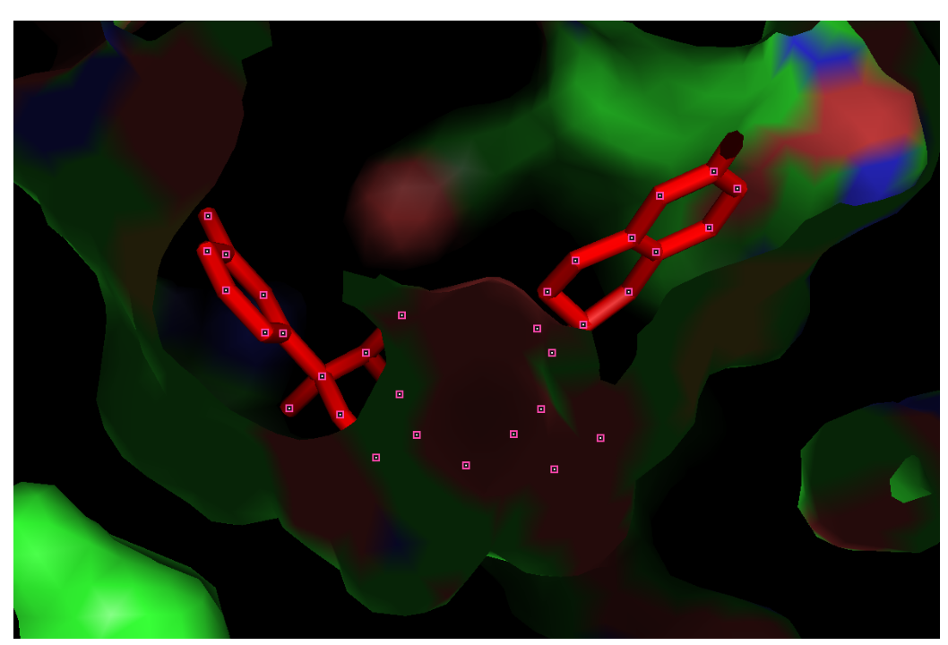
Figure 6: Structure of the ligand binding pocket from above.

JDTic interactions in the binding pocket illustrate selectivity
The JDTic antagonist, bound to the ligand binding pocket in the crystallographic structure of the kOR-JDTic complex, displayed high affinity interaction with the pocket residues (Ki=0.32nM), this property may also explain the potency and long duration of its binding as determined by structure-activity relationship assays (SARs). These studies also showed this ligand displays a 1000-fold selectivity for human k-OR as compared to other human opioid receptors. The interactions that comprise this selective JDTic-kOR interaction in the binding pocket have been characterized and comprise forms of polar, ionic and hydrophobic interactions with the receptor.
The interactions that stabilize JDTic binding to the k-OR pocket occur through 22 residues lining the pocket wall, which provide the contacts for these interactions to occur, all of them within 4.5Å of the ligand. These include salt bridges formed between the conserved Asp 138 residue at the bottom of the pocket and the protonated amines in both piperidine and isoquinoline moieties in the ligand. These two amino groups will anchor the ligand to the bottom of the cleft, making it acquire a V-shaped structure. The crystal structure of the JDTic complex also revealed that the ligand is partly stabilized though water-mediated polar interactions involving the distal hydroxyl groups in the piperidine and isoquinoline moieties (this data is confirmed by other relevant SAR studies). It was also noted that the majority of the residues lining the ligand binding pocket create a largely hydrophobic environment which is likely to play an important role in the stabilization of the aromatic rings in the two JDTic moieties. One of the most important of these hydrophobic interactions between the ligand and the binding pocket involved a conserved Trp287 and the ligand’s isopropyl group (it is thought this conserved amino acid residue plays an important role in blocking the activating conformational changes in class A GPCRs). Of all the numerous stabilizing interactions that are involved in the binding of the JDTic ligand to the k-OR pocket, arguably the most significant ones are those occurring in four residues which differ even between closely related opioid receptors. These residues are Val 108, Val 118, Ile 294 and Tyr 312 and are likely to play an important role in the selectivity of JDTic ligand binding of k-ORs. The valine and isoleucine residues provide hydrophic stabilization of the ligand whilst the tyrosine residue provides a polar interaction with JDTic. Analysis of JDTic ligand structure alone and bound to k-OR pocket revealed a degree of flexibility in the conformations it can acquire thanks to the available bond rotations allowed by the planar groups. This flexibility allows the ligand to explore a number of different conformations that facilitates its passage through the narrow and deep k-OR cleft and also allows the formation of a V-structure to correctly align the molecule in the pocket and stabilize its interactions. See some of the stabilizing interactions in Figure 7.
Figure 7. Some important molecular interactions between JDTic ligand and the binding pocket lining residues. Note the polar contacts with Asp138 and the hydrogen bonds involving the terminal hydroxyl groups (seem to disappear but are actually water-mediated). Note the Val 118 and Tyr312 residues which form characteristic interactions with the JDTic ligand and Trp287 which provides hydrophobic stabilisation of the ligand. Also labelled are Ile294 which is involved in forming important hydrophobic interactions with nor-BNI and GNTI morphine analogues and Tyr139 which will also form a hydrogen bond with both of these morphine analogues.

This particular image really shows the interactions between ligand and receptor well. The polar interactions shown give us a sense of the specificity of the ligand for the binding pocket and illustrates its affinity.
ReplyDelete